According to Rense.com: “Royal Raymond Rife was a brilliant scientist born in 1888 and died in 1971. After studying at Johns Hopkins, Rife developed technology which is still commonly used today in the fields of optics, electronics, radiochemistry, biochemistry, ballistics, and aviation. It is a fair statement that Rife practically developed bioelectric medicine himself.
He received 14 major awards and honors and was given an honorary Doctorate by the University of Heidelberg for his work. During the 66 years that Rife spent designing and building medical instruments, he worked for Zeiss Optics, the U.S. Government, and several private benefactors. Most notable was millionaire Henry Timkin, of Timkin roller bearing fame.” http://rense.com/health/rife.htm
Rife it seems was a shy and unassuming character and one cannot help but wonder how he came to become the charlatan described by Wiki below?
Wiki says: Rife’s claims could not be independently replicated,[5] and were discredited by independent researchers during the 1950s. Rife blamed the scientific rejection of his claims on a conspiracy involving the American Medical Association (AMA), the Department of Public Health, and other elements of “organized medicine”, which had “brainwashed and intimidated” his colleagues.[6]
(Wiki editors can’t believe that those who make lots of money and who are about to lose lots of money are capable of colluding in a conspiracy? Well, this is exactly what happened and it can be verified if they take the trouble to read a little history. )
…Interest in Rife’s claims was revived in some alternative medical circles by the 1987 book The Cancer Cure That Worked, which claimed that Rife had succeeded in curing cancer, but that his work was suppressed by a powerful conspiracy headed by the AMA… (Again, it was suppressed like many other things but Wiki editors call establishment suppression “conspiracy theory”.) https://en.wikipedia.org/wiki/Royal_Rife
It’s not that there is no history of Rife for the Wiki editors to use, I’ve listed numerous sources below. The story of Royal Raymond Rife is not unique in the annals of invention, cures and the work of brilliant minds. What happened to him and what became of his microscope is repeated again and again in the history of institutionalised scientific or medical denial and pseudo-debunking. Names like Gaston Naessens, Wilhelm Reich, Nikola Tesla and Immanuel Velikovsky come to mind as recipients of the full discrediting treatment at the hands of scientists and medical authorities. Scientists and doctors more interested in job security, money and prestige than serving humanity, as we are told they do. Rife’s crime arises not from mistakes, his microscope worked fine, his medical treatments have acclamation from the top names of the day. His unwitting crime was revolutionary; to cast doubt on germ theory, a theory that supports Big Pharma, an industry today worth billions that donates funding for ‘scientific research’ and he cured many incurable diseases, threatening the big medical cash cow. This is a story that surfaces so often as to become monotonous. Detectives advise, ‘follow the money’.
According to the Wiki oversimplification ‘Big Pharma conspiracy theory‘: “The term Big Pharma is used to refer collectively to global pharmaceutical industry.[2] According to Steve Novella the term has come to connote a demonised form of the pharmaceutical industry.[3] Professor of writing Robert Blaskiewicz has written that conspiracy theorists use the term Big Pharma as “shorthand for an abstract entity comprised of corporations, regulators, NGOs, politicians, and often physicians, all with a finger in the trillion-dollar prescription pharmaceutical pie”.” It fails to mention scientists.
Wiki again with ‘Luddite‘ a word that describes those who destroy machinery that is seen to threaten someones livelihood as it did the AMA: “The British Army clashed with the Luddites on several occasions. At one time, more British soldiers were fighting the Luddites than were fighting Napoleon on the Iberian Peninsula.” The Luddites were trying to keep their jobs and generations of skills just like the doctors and scientists of Rife’s day. The difference being that the scientists had the full support of the establishment and no soldiers came to protect the Rife microscope. This, in spite of the proven fact that it would save countless lives threatened by disease. It was all about scientific prestige and jobs, it’s something that has happened countless times both historically and more recently.
 No one knows what happened to the microscope or Rife’s notes. It’s like Deja Vu, the same thing happened to Gaston Naessens, Nikola Tesla’s notes disappeared just like John Keely‘s notes disappeared, all the books of Wilhelm Reich were burned. If you ask a scientists, and I have done so, about the names above they will, to a man (or woman) say they were all charlatans and pseudo scientists, but who would go to the trouble of removing every scrap of the life’s work of a charlatan? Any one who says there is not a concerted effort by science to cover its own mistakes is a fool and a nave. The mistake here is germ theory and there are a host of others.
Links and documents:
The reader needs to read the following links to understand what went on at the time:
http://rense.com/health/rife.htm
history_rife_cancer_treatment
An argument about Rife and about what constitutes evidence, among Wiki editors can be found here: wiki/Talk:Royal_Rife
Some video links:
[ http://www.youtube.com/watch?v=rvU9JrWycFI ]
[ http://www.youtube.com/watch?v=bM6jLwhdQEM ]
Timeline: Royal Raymond Rife
With many thanks to: http://educate-yourself.org/cn/rifetimelinemay1998.shtml
Compiled by A. Walter
May 1998
The original source seems to be: http://www.aceguru.com/Health/rife.htm
[Note from A. Walter: He (Rife) discovered an effective cure for cancer and many other diseases. Was he the greatest hero of the century? Read on and see what happened. ]
1888 Rife born in Elkhorn, Nebraska.
1913 Married. Moves to San Diego. A man of varied interests: ballistics, racing auto constructions, optics and microscopy.
1915-1918. Serves in the Navy. Sent to investigate foreign laboratories by the U.S. Government.
1920 Begins to investigate the possibilities of electric treatment of diseases. Timken, owner of Timken Roller Bearing Co., and Bridges of Bridges Carriage Co., provide funds to establish a laboratory and to finance his research. Begins research on tuberculosis.
1922 Begins cancer research.
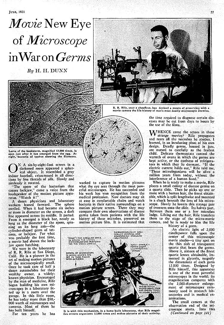
1929 October. Stock market crash. Two weeks later, Nov. 3, 1929, the San Diego Union carries an article announcing that Rife has built a microscope capable of staining living viruses with light to make them visible! (Today this is called bioluminescence.)
1931 Two men join Rife in his work: Dr. Arthur I. Kendall, Director of Medical Research at Northwestern University and Dr. Milbank Johnson of Pasadena Hospital. Dr. Alvin G. Frood, President of the American Association of Pathologists also becomes active in Rife’s research.
1931 Nov. 22: L.A. Times announces the discovery of a “filterable typhoid bacillus” being light-stained and observed to “change back into non- filterable form”, as seen through a powerful new microscope developed by Royal Raymond Rife that could directly observe living bacterium and viruses.
1932 May 3 & 4. Kendall speaks before the Assoc. of American Physicians at Johns Hopkins University telling of the preliminary successes with Rife’s methods and treatments. Dr. Thomas Rivers, virologist and bac-teriologist, Director of the Rockefeller Institute — a primary source of funding for medical research — and Dr. Hans Zinsser, call Kendall a liar to his face in front of the assembled crowd.
1932 July 5-7. Dr. Edward C. Rosenow of the Mayo Clinic’s Division of Experimental Bacteriology witnesses Rife’s results and becomes a supporter.
1932 Nov. 30. Rife isolates the filtrable virus of carcinoma. “Angle of refraction (polarization) 12 3/16 degrees; length 1/15 micron; breadth 1/20 micron, color by chemical refraction red-purple.” Pleomorphism also established.
1932 By end of the year “Rife can destroy the typhus bacteria, the polio virus, the herpes virus, the cancer virus and other viruses in culture and in experimental animals.”
1933 Rife completes the “universal microscope.” A resolution of 31,000 times and a magnification of 60,000 times.
1933 July. Dr. Karl Meyer, Director of the Hooper Foundation for Medical Research of UCSF joins Rife’s team.
1934 Summer: The first cancer clinic using Rife technology. A special University of Southern California Medical Research Committee chaired by Milbank Johnson is formed to oversee the research. Committee members are: Whalen Morrison, Chief Surgeon of the Santa Fe Railway. George C. Dock, M.D. George C. Fischer, M.D., Children’s Hospital of New York, Arthur I. Kendall, Dr. Zite, M.D., professor of pathology of Chicago University. Rufus B. Von Klein Schmidt, President of USC [University of Southern California].
Also in attendance:
Dr. James Couche of San Diego.
Dr. Carl Meyer, Ph.D. of the Hooper Foundation, SF.
Dr. Kopps of the Metabolic Clinic in La Jolla.
The clinic is held at the Scripps Institute in La Jolla, California. Sixteen terminally ill people are treated. Fourteen are cured in three months, the other two are cured in six months.
1935 Rife builds a smaller microscope that can be mass produced.
May – June: Dr. 0. Cameron Gruner of Montreal replicates Rife & Kendall’s cancer pleomorphism.
June: Four insurance companies are interested in financing Rife pro- vided the International Cancer Foundation gave its approval. Dr. Mildred Schram, Secretary of the Foundation, after visiting Rife’s lab, stipulates conditions for acceptance which have nothing to do with Rife’s work. Rife doesn’t have time to be sidetracked. Result: the Cancer Foundation never funds any of Rife’s work.
September: A new version of the Rife Frequency Ray is completed. October: Drs. Walker & Meyer of The Hooper Clinic, SF, using Rife’s microscope and “Beam Ray” replicate his cures.
November: Dr. Johnson opens a second clinic to test cures with the same incredible results as the first clinic.
1936 June 2: William Donner, President of the International Cancer Research Institute turns down Johnson’s application for research funds.
1937 July: Rife moves into his new lab on Alcott St. in Point Loma, built for him by his sponsor, Henry Timkin
1937 September – May: Johnson’s third clinic resulting in the same, identical cures. There is mounting pressure to go public. But Rife and his academic advisors, being cautious and recognizing the inevitable resistance from medical orthodoxy, are determined to gather as much irrefutable and massive statistical evidence as possible.
1937 Drs. Couche of San Diego, Gruner of Montreal are using Rifes Beam Ray with great success. In the fall Rife hires an engineer, Phil Hoyland, to help start a company for manufacturing the Beam Ray.
1938 The Rife Beam Ray Co. is in operation. Fourteen machines are built. Two go to England, one goes to Dr. Richard Hamer of the Paradise Valley Sanitarium, one to Dr. Arthur Yale, two to Arizona doctors, and eight to Southern California doctors. Dr. Hammer cures an 82-year-old from Chicago of terminal cancer. Through this man, Morris Fishbein, head of the AMA in Chicago learns of Rife and his work. Fishbein visits Rife. Wants to buy in. Rife and his associates turn him down.[This creates Fishbein’s prime revenge motivation for targeting Rife for indictment and the ultimate destruction of his work]
1938 The J.C Burnett Laboratory in New Jersy is burnt to the ground. Burnett’s wife is a member of the Timken family. The lab is burned while Burnett and his wife are visiting Rife!
The AMA indicts Rife for fraudulent medical practices.
1938 June 12: Opening day of Rife’s trial. During his testimony, Rife is so nervous he cannot stop shaking. A doctor recommends he take a drink to calm himself. Rife’s alcoholism begins.
During the trial and afterwards the AMA visits all doctors involved with Rife. “Those who didn’t stop using the Frequency Instruments lose their medical license. Dr. Hamer quickly returns his instrument. Dr. Gruner returns his. Dr. Couche defies the AMA and his license is re- voked. Many other doctors associated with Rife turn their backs on him including Drs. Drood, Rosenow, & Meyer. Rife nevertheless wins the case. The AMA pays off Kendall in Baltimore “about” $200,000. He goes to Mexico,
1940 Kendall dies.
1944 Milbank Johnson dies under suspicious circumstances. Two federal inspectors conclude that he was likely poisoned. Reputedly he was to present the long delayed Rife findings to the AMA the following day. All Rife’s records kept by his academic committee are destroyed. The committee disbands.
1946 Rife’s drinking forces him to sell off his lab piece by piece. He is committed for “alcohol rehabilitation.”
1948 Drs. Virginia Livingston-Wheeler & Eleanor Alexander-Jackson, micro- biologists in Philadelphia prove that the cancer virus “is in actuality a pleomorphic bacterium.”
1949 Dr. Virginia Livingston becomes the head of New Rutgers Presbyterian Laboratory in Newark, NJ.
1949 June 6: Morris Fishbein is ousted by the AMA at its Atlantic city convention. Reason: years of advertising fraud and fund stealing.
1950 Rife is released from rehabilitation and returns to work. He forms a partnership with John Crane, an engineer/scientist. Crane “re-invents” the Beam Ray and hires Verne Thomson, an electronics expert with the San Diego police force, to help construct the new machines.
Dr. Irene Corey Diller of the Institute for Cancer research in Philadelphia isolates fungus agents from cancer growths. Unknowingly she has replicated Rife and Gruner’s work. She sets up a symposium in New York in order to announce her discovery. It is killed by Dr. Cornelius P. Rhoads, the head of Memorial Sloan-Kettering Cancer Center.
Dr. James Hillman of RCA Labs in Princeton. NJ, later confirms the Livingston-Wheeler-Gruner-Rife pleomorphism.
1953 Dr. Diller publishes her discovery: Studies of Fungoid Form Found in Malignancy. Dr. Livingston-Wheeler presents her own discoveries to the 6th International Congress of Microbiology in Rome. Sep. 10: The Washington Post reports. “The New York Academy of Medicine immediately discounts the announcement.” Dr. Rhoads of Memorial Sloan-Kettering Cancer Center stops all funds for the Rutgers-Presbyterian Hospital Laboratory. The lab is closed, putting Dr. Livingston-Wheeler out of business.
1954 Dr. Livingston-Wheeler moves to San Diego, taking a job with a clinic. The Committee on Cancer Diagnosis and Therapy of the National Research Council “evaluate” Rife’s discoveries and conclude that “they couldn’t work.” (Please note: never at any point, and to this date, no orthodox cancer agency has tested Rife’s work.)
Rife, under Crane’s insistence, copyrights a description of his cancer cure.
1958 Jan: A group of Salt Lake City doctors begin using the Rife Frequency machine. In May the Salt Lake City Medical Board forces them to stop using it.
The California Public Health Dept. holds a hearing. A Frequency Instrument had been provided for testing to the Palo Alto Detection Lab., the Kalbfeld Lab., the UCLA Medical Lab. and the San Diego Testing Lab. All declared that it was safe to use. Result: The AMA Board under the Calif. Director of Public Health, Dr. Malcolm Merrill declares it unsafe and bans it from the market.
1958 July 14. Or Virginia Livingston-Wheeler is the first speaker at the 1st International Congress of Microbiology and Leukemia in Antwerp, Belgium. She discovers that the pleomorphism of cancer is widely accepted in Europe while ignored in the U.S.
1958 November: After six months of testing Rife’s technology, Dr. Robert Stafford of Dayton, Ohio, presents his findings to the Executive Committee of the General Practice Section of the Montgomery County Medical Society of the AMA. The committee is impressed. They set up a research committee from Dayton’s most influential doctors.
1959 Dr. Clara Fonti of Milan, Italy “innoculates herself with a bacterial culture of cancer. She grows a tumor, later surgically removed. Thereby proving pleomorphism as a factor in human cancer.
Dr. Livingston-Wheeler meets her neighbor Royal R. Rife.
1959-1960 Dr. Livingston & Rife work together. She arranges for the Institute of Cancer Research in Philadelphia to provide Rife with mice. Their view on pleomorphism are much the same. The only difference is that Dr. Livingston intends to develop a serum while Rife knows the virus dis-integrates under his Beam Ray.
1960 John Crane writes and copyrights a manual explaining how the Frequency Instrument is to be used. Dr. Stafford of Dayton suggests that he, Stafford, manufacture and distribute the machine in the USA. Crane decides to license the machine to prevent doctors from changing it, thus failing to get results. Ninety machines are distributed “for research and verification on notorized contracts.”
1960 The AMA and the FDA strike. Crane’s office is raided. $40,000 in equipment is taken along with all engineering data, research records and reports, pictures off the wall, private letters, invoices, tape recordings, electronic parts. All without a search warrant! Doctors who have the machines are visited and forced to give them up. Ordinary citizens who have begun experimenting personally are threatened. One woman is hospitalized from shock by the AMA raid.
Rife and Crane are arrested and released on bail. Rife, almost 73, unable to handle more abuse, goes into hiding in Mexico.
1961 Spring. John Crane’s trial lasts 24 days. “The records and materials seized are not allowed to be used by Crane in his own defense. . . Rife’s deposition is not permitted to be introduced.” The foreman of the jury is an AMA doctor; the balance of the jury is screened to make sure they have no medical nor electronic knowledge. “No medical reports from the 30’s and 40’s are admitted. Neither are other doctor’s reports. Nor is a Frequency Instrument demonstrated much less admitted into court. “The only medical opinion offered by the State of California is from Dr. Paul Shae who had been given a Frequency Instrument by the Public Health Department 2 months before the trial. Shea admits he never tried (it) or made tests to evaluate it. He simply examined it and decided that it had no curative powers and didn’t lend itself to investigative use”.
Crane is found guilty and sentenced to 10 years in prison. Later the State Supreme Court, on appeal, reverses two of the three counts against Crane “because no specific criminal intent had been proven.” Crane spends three years and one month in jail. After Crane’s imprisonment Dr. Stafford of Dayton is forced to give up his instrument and to give up medicine. A Salt Lake City Doctor’s instrument was sabotaged and he was so hounded by the orthodox medical authorities that he commits suicide.
1962 Dr. Livingston has a heart attack. She recovers.
Kefauver amendments to the Food and Drug Act of 1938 grant to the FDA the right to determine if a drug is effective. Safety is no longer a prime consideration. Drug treatment effectiveness and safety is there-by taken out of the hands of the doctor and his patient.
1964 John Crane is released from prison. He begins the fight all over again. 1965 October: Crane submits an application to the Calif. Board of Public
Health for approval of the Frequency Instrument. “The application is made in the name of Rife Microscope Institute,” John Crane, owner. The Health Department answers that Crane must first show the instrument to be effective.
Dr. Charles W. Bunner, Chiropractor, agrees to provide “proof of effectiveness.” The Calif. Department of Health pays him a visit and forbids him to use the instrument, and present him with a court order to have it destroyed.
Dr. Les Drown, Chiropractor, provides a statement. An American Cancer Society representative subsequently forces him to “sign over” his Frequency Instrument or go to jail. (Rife’s cancer discoveries are never patented.) Rife returns from Mexico.
1966 Dr. Livingston and her old colleague, Dr. Eleanor Alexander-Jackson present a paper at an American Cancer Seminar in Arizona. When Dr. Alexander-Jackson returns to Columbia University she discovers that she and her work have been terminated.
1968 Dr. Livingston and her husband open a clinic in San Diego.
1968-1983 They treat over 10,000 cancer patients utilizing her serum and a high immune-building diet. An 80-percent success rate. (The State of California Department of Health has subsequently tried to shut down her clinic. They have also outlawed the use of her serum. —Personal communication from the Livingston Medical Center.)
1969 March 4: Rife signs ownership of his microscope over to John F. Crane. It is Crane who preserves all of Rife’s work that remains.
1969 Nov. 5-8: Drs. Livingston, Alexander, Diller and Dr. Florence Seibert from the Veterans Administration Research Laboratory in Bay Pines, Florida present a paper: Microorganisms Associated with Malignancy.
1970 Oct. 30: Their paper is published.
1970 Rife dies. He has been hospitalized for intoxication. Records show he is given an extra (overdose) of Valium. A mixture of Valium and alcohol is lethal. He is 84.
1971 December 23: President Richard Nixon signs a $1.6 billion law to open the “war on cancer.”
1972 Dr. Livingston publishes her first book: Cancer: A Breakthrough. She condemns the NCI for its misuse of money and “the use of people as guinea pigs for a ‘surgery-radiation-chemotherapy’ program dictated by special interests.”
1973 The Supreme Court rules that the FDA “can decide without a hearing which evidence it would allow.”
1980 “The AMA is found guilty by a U.S. Court of Appeals of ‘conspiracy to restrain competition. . . New methods of health care have been discouraged, restricted and in some instances eliminated.”‘
1985 The Sloan-Kettering Cancer Institute finds the Rife-Livingstone-etc. organism (virus) in all blood cultures of cancer patients. They conclude that the organism comes from outside contamination and bury the report.
1984 Dr. Livingston publishes The Conquest of Cancer.
1985 By this time the National Cancer Institute is spending $1.2 billion dollars annually for cancer. This does not count the monies raised by the American Cancer Society.
1988 Rife Labs is formed to revitalize Rife’s work.
1990 It is estimated that $50 billion dollars have been spent on the “War on Cancer.” Twenty percent of this money is spent on actual research. Dr. Virginia Livingston-Wheeler dies.
1996 June: John Crane dies totally destitute in San Diego County. (Personal communication from the Cancer Research Organization.)
1998 May: Nothing has changed as of this writing. Rife Frequency Treatments are illegal except for experimental purposes. It reputedly cures AIDS but the frequencies are absolutely forbidden to be used even for experimental purposes in the United States only. The existence of the Black Field microscope continues to be denied by orthodox medicine as is the pleomorphism of bacteria/viruses. The suppression of alternative cancer treatments remain in full force and now also includes Hydrazine Sulfate.
Just last year the FDA raided and demolished the San Diego offices of American Biologics whose clinic in Tijuana uses alternate cancer treatments.
How long can this last?
Compiled by A. Walter, 5/98
Morris Fishbein – AMA Enemy Of American Health
From the Annual Report of the Board of Regents of
THE SMITHSONIAN INSTITUTION – 1944
The Universal Microscope
Note: because important web-sites are frequently “here today but gone tomorrow”, the following was archived from http://www.keelynet.com/biology/rife2.txt on January 31, 2003. . This is NOT an attempt to divert readers from the aforementioned web-site. Indeed, the reader should only read this back-up copy if it cannot be found at the original author’s site.
archived as http://www.stealthskater.com/Documents/Rife_1.doc
11/26/89 RIFE2.ASC
It is only a reasonable supposition; but already in one instance, a very successful and highly commendable achievement on the part of of San Diego, California, who, for many years, has built and worked with light microscopes which far surpasses the theoretical limitations of the ordinary variety of instrument, all the Rife scopes possessing superior ability to attain high magnification with accompanying high resolution.
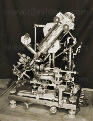
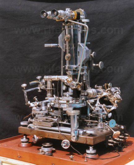
The largest and most powerful of these, the Universal Microscope, developed in 1933, consists of 5,682 parts and is so called because of its adaptability in all fields of microscopical work, being fully equipped with separate substage condenser units for transmitted and monochromatic beam dark-field, polarized, and slit-ultra illumination, including also a special device for crystallography. The entire optical system of lenses and prisms as well as the illuminating units are made of block-crystal quartz, quartz being especially transparent to ultraviolet radiations.
This illuminating unit used for examining the filterable forms of disease organisms contains 14 lenses and prisms, 3 of which are in the high-intensity incandescent lamp, 4 in the Risley prism, and 7 in the achromatic condenser which, incidentally, has a numerical aperture of 1.40. Between the source of light and the specimen are subtended two circular, wedge-shaped, block-crystal quartz prisms for the purpose of polarizing the light passing through the specimen, polarization being the practical application of the theory that light waves vibrate in all planes perpendicular to the direction in which they are propagated.
Therefore, when light comes into contact with a polarizing prism, it is divided or split into two beams, one of which is refracted to such an extent that it is reflected to the side of the prism without, of course, passing through the prism while the second ray, bent considerably less, is thus enabled to pass through the prism to illuminate the specimen.
When the quartz prisms on the universal microscope, which may be rotated with vernier control through 360 degrees, are rotated in opposite directions, they serve to bend the transmitted beams of light at variable angles of incidence while, at the same time, a spectrum is projected up into the axis of the microscope, or rather a small portion of the spectrum to the other, going all the way from the infrared to the ultraviolet.
NOW, WHEN THAT PORTION OF THE SPECTRUM IS REACHED IN WHICH BOTH THE ORGANISM AND THE COLOR BAND VIBRATE IN EXACT ACCORD, ONE WITH THE OTHER, A DEFINITE CHARACTERISTIC SPECTRUM IS EMITTED BY THE ORGANISM.
In the case of the filter-passing form of the BACILLUS TYPHOSUS, for instance, A BLUE SPECTRUM IS EMITTED AND THE PLANE OF POLARIZATION DEVIATED PLUS (+) 4.8 DEGREES.
The predominating chemical constituents of the organism are next ascertained after which the quartz prisms are adjusted or set, by means of vernier control, to minus (-) 4.8 degrees (again in the case of the filter-passing form of the BACILLUS TYPHOSUS) so that the opposite angle of refraction may be obtained.
A MONOCHROMATIC BEAM OF LIGHT, CORRESPONDING **EXACTLY** TO THE FREQUENCY OF THE ORGANISM (for Dr. Rife has found that EACH DISEASE ORGANISM RESPONDS TO AND HAS A DEFINITE AND DISTINCT WAVE LENGTH, a fact confirmed by British medical research workers) IS THEN SENT UP THROUGH THE SPECIMEN AND THE DIRECT TRANSMITTED LIGHT, THUS ENABLING THE OBSERVER TO VIEW THE ORGANISM STAINED IN ITS TRUE CHEMICAL COLOR and revealing ITS OWN INDIVIDUAL STRUCTURE IN A FIELD WHICH IS BRILLIANT WITH LIGHT.
The objectives used on the universal microscope are a 1.12 dry lens, a 1.16 water immersion, a 1.18 oil immersion, and a 1.25 oil immersion. The rays of light refracted by the specimen enter the objective and are then carried up the tube IN PARALLEL RAYS through 21 light bends to the ocular, A TOLERANCE OF LESS THAN ONE WAVE LENGTH OF VISIBLE LIGHT ONLY BEING PERMITTED N THE CORE BEAM, OR CHIEF RAY, OF ILLUMINATION.
Now, instead of the light rays starting up the tube in a parallel fashion, TENDING TO CONVERGE AS THEY RISE HIGHER AND FINALLY CROSSING EACH OTHER, arriving at the ocular SEPARATED BY CONSIDERABLE DISTANCE as would be the case with an ordinary microscope, IN THE UNIVERSAL TUBE THE RAYS ALSO START THEIR RISE PARALLEL TO EACH OTHER BUT, JUST AS THEY ARE ABOUT TO PULL THEM OUT PARALLEL AGAIN, ANOTHER PRISM BEING INSERTED EACH TIME THE RAYS ARE ABOUT READY TO CROSS.
These prisms, inserted in the tube, which are adjusted and held in alignment by micrometer screws of 100 threads to the inch in special tracks made of magnelium (magnelium having the closest coefficient of expansion of any metal to quartz), are separated by a distance OF ONLY 30 MILLIMETERS.
Thus, THE GREATEST DISTANCE THAT THE IMAGE in the universal microscope IS PROJECTED THROUGH ANY ONE MEDIA, EITHER QUARTZ OR AIR, IS 30 MILLIMETERS INSTEAD OF THE 160, 180, OR 190 MILLIMETERS as in the empty or air-filled tubes of an ordinary microscope, the total distance which the light rays travel ZIGZAG FASHION through the universal tube being 449 MILLIMETERS, although the physical length of the tube itself is 229 millimeters.
It will be recalled that if one pierces a black strip of paper or cardboard with the point of a needle and then brings the card up close to the eye so that the hole is in the optic axis, a small brilliantly lighted object will appear LARGER AND CLEARER, REVEALING MORE FINE DETAIL, than if it were viewed from the same distance without the assistance of the card.
This is explained by the fact that the beam of light passing through the card is very narrow, the rays entering the eye, therefore, being practically parallel, whereas without the card the beam of light is much wider and the DIFFUSION CIRCLES MUCH LARGER. It is this principle of parallel rays in the universal microscope and the resultant shortening of projection distance between any two blocks or prisms plus the fact that objectives can thus be substituted for oculars, these “oculars” being THREE MATCHED PAIRS OF 10-MILLIMETER, 7-MILLIMETER, AND 4-MILLIMETER OBJECTIVES IN SHORT MOUNTS, which would make possible not only the unusually high magnification and resolution but which SERVE TO ELIMINATE ALL DISTORTION AS WELL AS ALL CHROMATIC AND SPHERICAL ABERRATION.
Quartz slides with especially thin quartz cover glasses are used when a tissue section or culture slant is examined, the tissue section itself also being very thin. An additional observational tube and ocular which yield a magnification of 1,800 diameters are provided so that that portion of the specimen which it is desired should be examined may be located and so that the observer can adjust himself more readily when viewing a section at a high magnification.
The universal stage is a double rotating stage graduated through 360 degrees in quarter-minute divisions, the upper segment carrying the mechanical stage having a movement of 40 degrees, plus or minus. Heavily constructed joints and screw adjustments maintain rigidity of the microscope which weighs 200 pounds and stands 24 inches high, the bases of the scope being nickel-cast steel plates, accurately surfaced, and equipped with three leveling screws and two spirit levels set at angles of 90 degrees. The coarse adjustment, a block thread screw with 40 threads to the inch, slides in a 1 1/2 dovetail which gibes directly onto the pillar post. The weight of the quadruple nosepiece and the objective system is taken care of by the intermediate adjustment at the top of the body tube. The stage, in conjunction with a hydraulic life, acts as a lever in operating the fine adjustment. A 6-gauge screw having 100 threads to the inch is worked through a gland into a hollow, glycerine-filled post, the glycerine being displaced and replaced at will as the screw is turned clockwise or anticlockwise, allowing a 5-to-1 ratio on the lead screw. This, accordingly, assures complete absence of drag and inertia. The fine adjustment being 700 times more sensitive then that of ordinary microscopes, the length of time required to focus the universal ranges up to 1-1/2 hours which, while on first consideration, may seem a disadvantage, is after all but a slight inconvenience when compared with the many years of research and the hundreds of thousands of dollars spent and being spent in an effort to isolate and to look upon disease-causing organisms in their true form.
Working together back in 1931 and using one of the smaller Rife microscope having a magnification and resolution of 17,000 diameters, Dr. Rife and Dr. Arthur Isaac Kendall of the department of bacteriology of Northwestern University Medical School were able to observe and demonstrate the presence of the filter-passing forms of BACILLUS TYPHOSUS. An agar slant culture of the Rawlings strain of BACILLUS TYPHOSUS was first prepared by Dr. Kendall and inoculated into 6 cc. of “Kendall” K Medium, a medium rich in protein but poor in peptone and consisting of 100 mg. of dries hog intestine and 6 cc. of tyrode solution (containing neither glucose nor glycerine) which mixture is shaken well so as to moisten the dried intestine powder and then sterilized in the autoclave, 15 pounds for 15 minutes, alterations of the medium being frequently necessary depending upon the requirements for different organisms. Now, after a period of 18 hours in this K Medium, the culture was passed through a Berkefeld “N” filter, a drop of the filtrate being added to another 6 cc. of K Medium and incubated ar 37 degrees C. Forty-eight hours later this same process was repeated, the “N” filter again being used, after which it was noted that the culture no longer responded to peptone medium, growing now only in the protein medium. When again, within 24 hours, the culture was passed through a filter-the finest Berkefeld “W” filter, a drop of the filtrate was once more added to 6 cc. of K Medium and incubated at 37 degrees C., a period of 3 days elapsing before a new culture was transferred to K Medium and yet another 3 days before a new culture was prepared. Then, viewed under an ordinary microscope, these cultures were observed to be turbid and to reveal no bacilli whatsoever. When viewed by means of dark-field illumination and oil-immersion lens, however, the presence of small, actively motile granules was established, although nothing at all of their individual structure could be ascertained. Another period of 4 days was allowed to elapse before these cultures were transferred to K Medium and incubated at 37 degrees C. for 24 hours when they were then examined under the Rife microscope where, as was mentioned earlier, the filterable typhoid bacilli, emitting a blue spectrum, caused the plane of polarization to be deviated plus 4.8 degrees. Then when the opposite angle of refraction was obtained by means of adjusting the polarizing prisms to minus 4.8 degrees and the cultures illuminated by a monochromatic beam coordinated in frequency with the chemical constituents of the typhoid bacillus, small oval actively motile, bright turquoise-blue bodies were observed at a magnification of 5,000 diameters, in high contrast to the colorless and motionless debris of the medium. These observations were repeated eight times, the complete absence of these bodies in uninoculated control K Media also being noted.
To further confirm their findings, Drs. Rife and Kendall nest examined 18-hour-old cultures which had been inoculated into K Medium and incubated at 37 degrees C., since it is just at this stage of growth in this medium and at this temperature that the cultures become filterable. And, just as had been anticipated, ordinary dark-field examination revealed unchanged, long, actively motile bacilli; bacilli having granules within their substance; and free-swimming, actively motile granules; while under the Rife microscope were demonstrated the same long, unchanged, almost colorless bacilli; bacilli, practically colorless, inside and at one end of which was a turquoise-blue granule resembling the filterable forms of the typhoid bacillus; and free-swimming, small, oval, actively motile, turquoise-blue granules. By transplanting the cultures of the filter-passing organisms or virus into a broth, they were seen to change over again into their original rod-like forms.
At the same time that these findings of Drs. Rife and Kendall were confirmed by Dr. Edward C. Rosenow of the Mayo Foundation, the magnification with accompanying resolution of 8,000 diameters of the Rife microscope, operated by Dr. Rife, was checked against a dark-field oil-immersion scope operated by Dr. Kendall and an ordinary 2-mm. oil-immersion objective, x10 ocular, Zeiss scope operated by Dr. Rosenow at a magnification of 900 diameters. Examinations of gram and safranin-stained films of culture of Bacillus typhosus, gram and safranin-stained films of blood and of the sediment of the spinal fluid from a case of acute poliomyelitis were made with the result that bacilli, streptococci, erythrocytes, polymorphonuclear leukocytes, and lymphocytes measuring nine times the diameter of the same specimens observed under the Zeiss scope at a magnification and resolution of 900 diameters, were revealed with unusual clarity. Seem under the dark-field microscope were moving bodies presumed to be the filterable turquois-blue bodies of the typhoid bacillus which, as Dr. Rosenow has declared in his report (Observations on filter-passing forms of Eberthella-typhi-Bacillus typhosus – and of the streptococcus from poliomyelitis, Proc. Staff Meeting Mayo Clinic, July 13, 1932), were so “unmistakably demonstrated” with Rife microscope, while under the Zeiss scope stained and hanging-drop preparations of clouded filtrate culture were found to be uniformly negative. With the Rife microscope also were demonstrated brownish-gray cocci and diplococci in hanging-drop preparations of the filtrates of streptococcus from poliomyelitis. These cocci and diplococci, similar in size and shape to those seen in the culture although of more uniform intensity, and characteristic of the medium in which they had been cultivated, were surrounded by a clear halo about twice the width of that at the margins of the debris and of the Bacillus typhosus. Stained films of filtrates and filtrate sediments examined under the Zeiss microscope, and hanging-drop, dark-field preparations revealed no organisms, however. Brownish-gray cocci and diplococci of the exact same size and density as those observed in the filtrates of the streptococcus cultures were also revealed in hanging-drop preparations of the virus of poliomyelitis under the Rife microscope, while no organisms at all could be seen in either the stained films of filtrates and filtrate sediments examined with the Zeiss scope or in hanging-drop preparations examined by means of the dark-field. Again using the Rife microscope at a magnification of 8,000 diameters, numerous nonmotile cocci and diplococci of a bright-to-pale pink in color were seen in hanging-drop preparations of filtrates of Herpes encephalitic virus.
Although these were observed to be comparatively smaller then the cocci and diplococci of the streptococcus and poliomyelitis viruses, they were shown to be of fairly even density, size and form and surrounded by a halo. Again, both the dark-field and Zeiss scopes failed to reveal any organisms, and none of the three microscopes disclosed the presence of such diplococci in hanging-drop preparation of the filtrate of a normal rabbit brain. Dr. Rosenow has since revealed these organisms with the ordinary microscope at a magnification of 1,000 diameters by means of his special staining method and with the electron microscope at a magnification of 12,000 diameters. Dr. Rosenow has expressed the opinion that the inability to see these and other similarly revealed organisms is due, not necessarily to the minuteness of the organisms, but rather to the fact that they are of a nonstaining, hyaline structure. Results with the Rife microscopes, he thinks, are due to the “ingenious methods employed rather than to excessively high magnification.” He has declared also, in the report mentioned previously, that “Examination under the Rife microscope of specimens containing objects visible with the ordinary microscope, leaves no doubt of the accurate visualization of objects or particulate matter by direct observation at the extremely high magnification obtained with this instrument.”
Exceedingly high powers of magnification with accompanying high powers of resolution may be realized with all of the Rife microscopes, one of which, having magnification and resolution up to 18,000 diameters, is now being used at the British School of Tropical Medicine in England. In a recent demonstration of another of the smaller Rife scopes (May 16,1942) before a group of doctors including Dr. J.H.Renner of Santa Barbara, Calif.; Dr. Roger A. Schmidt of San Francisco, Calif.; Dr. Lois Bronson Slade of Alameda, Calif.; Dr.Lucile B. Larkin of Bellingham, Wash.; Dr. E. F. Larki, of Bellingham, Wash.; and Dr. W. J. Gier, of San Diego, Calif., a Zeiss ruled grading was examined, first under an ordinary commercial microscope equipped with a 1.8 high dry lens and X 10 ocular, and then under the Rife microscope. Whereas 50 lines were revealed with the commercial instrument and considerable aberration, both chromatic and spherical noted, only 5 lines were seen with the Rife scope, these 5 lines being so highly magnified that they occupied the entire field, without any aberration whatsoever being apparent. Dr. Renner, in a discussion of his observations, stated that “The entire field to its very edges and across the center had a uniform clearness that was not true on the conventional instrument.” Following the examination of the grading, an ordinary unstained blood film was observed under the same two microscopes. In this instance, 100 cells were seen to spread throughout the field of the commercial instrument while but 10 cells filled the field of the Rife scope.
The universal microscope, of course, is the most powerful Rife scope, possessing a resolution of 31,000 diameters and magnification of 60,000 diameters. With this it is possible to view the interior of the `pin-point’ cells, those cells situated between the normal tissue cells and just visible under the ordinary microscope, and to observe the smaller cells which compose the interior of these pin-point cells. When one of these smaller cells in magnified, still smaller cells are seen within its structure. And when one of the still smaller cells, in its turn, is magnified, it, too, is seen to be composed of smaller cells. Each of the 16 times this process of magnification and resolution can be repeated, it is demonstrated that there are smaller cells within the smaller cells, a fact which amply testifies as to the magnification and resolving power obtainable with the universal microscope.
More then 20,000 laboratory cultures of carcinoma were grown and studied over a period of 7 years by Dr. Rife and his assistants in what, at the time, appeared to be a fruitless effort to isolate the filter-passing form, or virus, which Dr. Rife believed to be present in this condition. Then in 1932, the reactions in growth of bacterial cultures to light from the rare gasses were observed, indicating a new approach to the problem. Accordingly, blocks of tissue one-half centimeter square, taken from an unulcerated breast carcinoma, were placed in a circular glass loop filled with argon gas to a pressure of 14 millimeters, and a current of 5,000 volts applied for 24 hours, after which the tubes were placed in a 2-inch water vacuum and incubated at 37 degrees C. for 24 hours. Using a specially designed 1.12 dry lens, equal in amplitude of magnification to the 2-mm. apochromatic oil-immersion lens, the cultures wee then examined under the universal microscope, at a magnification of 10,000 diameters, where very much animated, purplish-red, filterable forms, measuring less then one-twentieth of a micron in dimension, were observed. Carried through 14 transplants from K Medium to K Medium, this B. X. virus remained constant; inoculated into 426 Albino rats, tumors `with all the true pathology of neoplastic tissue’ were developed. Experiments conducted in the Rife Laboratories have established the fact that these characteristic diplococci are found in the blood monocytes in 92 percent of all cases of neoplastic diseases. It has also been demonstrated that the virus of cancer, like the viruses of other diseases, can be easily changed from one form to another by means of altering the media upon which it is grown. With the first change in media, the B. X. virus becomes considerable enlarged although its purplish-red color remains unchanged. Observation of the organism with an ordinary microscope is made possible by a second alteration of the media. A third change is undergone upon asparagus base media where the B. X. virus is transformed from its filterable state into cryptomyces pleomorphia fungi, these fungi being identical morphologically both microscopically to that of the orchid and of the mushroom. And yet a fourth change may be said to take place when this cryptomyces pleomorphia, permitted to stand as a stock culture for the period of metastasis, becomes the well-known mahogany-colored Bacillus coli.
It is Dr. Rife’s belief that all micro-organisms fall into 1 of not more then 10 individual groups (Dr. Rosenow has stated that some of the viruses belong to the group of the streptococcus), and that any alteration of artificial media of slight metabolic variation in tissues will induce an organism of one group to change over into any other organism included in that same group, it being possible, incidentally, to carry such changes in media or tissues to the point where the organisms fail to respond to standard laboratory methods of diagnosis. These changes can be made to take place in as short a period of time as 48 hours. For instance, by altering the media – 4 parts per million per volume – the pure culture of mahogany- colored Bacillus coli becomes the turquoise-blue Bacillus typhosus. Viruses of primordial cells of organisms which would ordinarily require an 8-week incubation period to attain their filterable state, have been shown to produce disease within 3 days’ time, proving Dr. Rife’s contention that the incubation period of a micro-organism is really only a cycle of reversion.
He states: “In reality, it is not the bacteria themselves that produce the disease, but we believe the chemical constituents of these micro-organisms enacting upon the unbalanced cell metabolism of the human body that in actuality produce the disease. We also believe if the metabolism of the human body is perfectly balanced or poised, it is susceptible to no disease.”
In other word, the human body itself is chemical in nature, being comprised of many chemical elements which provide the media upon which the wealth of bacteria normally present in the human system feed. These bacteria are able to reproduce. They too are composed of chemicals. Therefore, if the media upon which they feed, in this instance the chemicals or some portion of the chemicals of the human body, become changed from the normal, it stands to reason that these same bacteria, or at least certain numbers of them, will also undergo a change chemically since they are now feeding upon media which are not normal to them, perhaps being supplied with too much or too little of what they need to maintain a normal existence. They change, passing usually through several stages of growth, emerging finally as some entirely new entity – as different morphologically as are the caterpillar and the butterfly (to use an illustration given us). The majority of the viruses have been definitely revealed as living organisms, foreign organisms it is true, but which once were normal inhabitants of the human body -living entities of a chemical nature of composition.
Under the universal microscope disease organisms such as those of tuberculosis, cancer, sarcoma, streptococcus, typhoid, staphylococcus, leprosy, hoof and mouth disease, and others may be observed to succumb when exposed to certain lethal frequencies, coordinated with the particular frequencies peculiar to each individual organism, and directed upon them by rays covering a wide range of waves. By means of a camera attachment and a motion-picture camera not built into the instrument, many “still” micrographs as well as hundreds of feet of motion-picture film bear witness to the complete life cycles of numerous organisms. It should be emphasized, perhaps, that invariably the same organisms refract the same colors when stained by means of the monochromatic beam of illumination on the universal microscope, regardless of the media upon which they re grown. The virus of theBacillus typhosus is always a turquoise blue, the Bacillus coli always mahogany colored, the Mycobacterium leprae always a ruby shade, the filter-passing form of virus of tuberculosis always an emerald green, the virus of cancer always a purplish red, and so on. Thus, with the aid of this microscope, it is possible to reveal the typhoid organism, for instance, in the blood of a suspected typhoid patient 4 and 5 days before a Widal is positive. When it is desired to observe the flagella of the typhoid-organism, Hg salts are used as the medium to see at a magnification of 10,000 diameters.
In the light of the amazing results obtainable with this universal microscope and its smaller brother scopes, there can be no doubt of the ability of these instruments to actually reveal any and all microorganisms according to their individual structure and chemical constituents.
With the aid of its new eyes – the new microscopes, all of which are continually being improved – science has at last penetrated beyond the boundary of accepted theory and into the world of the viruses with the result that we can look forward to discovering new treatments and methods of combating the deadly organisms – for science dose not rest.
To Dr. Karl K. Darrow, Dr. John A. Kolmer, Dr. William P. Lang, Dr. L. Marton, Dr. J. H. Renner, Dr. Royal R. Rife, Dr. Edward C. Rosenow, Dr. Arthur W. Yale, and Dr. V. K. Zworykin,
we wish to express our appreciation for the help and information so kindly given us and to express our gratitude, also, for the interest shown in this effort of bringing to the attention of more of the medical profession the possibilities offered by the new microscopes.
DISCUSSION
OBSERVATIONS WITH THE RIFE MICROSCOPE
OF FILTER-PASSING FORMS OF MICROORGANISMS
Recently, I reported to the staff of the Mayo Clinic the more imported observation made during three days, July 5, 6 and 7, 1932, spent in Dr. Kendall’s laboratory at Northwestern University Medical School, Chicago. I went there at the invitation of Drs. Kendall and Rife to share with them their observations in a restudy of the filter-passing forms of Eberthella typhi as seen with an improved model of the Rife microscope. They asked me also to bring with me my cultures of the streptococcus from poliomyelitis.
I would like to repeat here that portion of my report which had to do specifically with the Rife microscope.
Owing to the novel and important character of the work, each of us verified at every step the results obtained. Microscopic examinations of suitable specimens were made as a routine by Dr. Rife with his high-power microscope, by Dr. Kendall with the oil immersion dark-field, and by myself with the ordinary Zeiss microscope equipped with a 2 mm apochromatic oil immersion lens and 100 X 10 ocular giving a magnification of about 90 diameters. Most observations with the Rife microscope were made at 8,000 diameters. In order to check the magnification, gram and safranin stained films of cultures of Eberthella typhi, of the streptococcus from poliomyelitis, and stained films of blood, and of the sediment of the spinal fluid from a case of acute poliomyelitis were examined. Bacilli, streptococci, erythrocytes, polymorphonuclear leukoeytes and lymphocytes were clearly seen, and in each instance were, as nearly as could be examined with the 2 mm oil immersion at about 900 diameters.
The following principles and methods were stated by Dr. Rife as being essential in order to visualize clearly the objects at this and higher magnifications by direct observation. Spherical aberration is reduced to the minimum and magnification greatly increased by using objectives in place of oculars. Proper visualization, especially of unstained objects, is obtained by the use of an intense beam of monochromatic polarizes light created by rotating wedge-shaped quartz prisms placed between the source of light and the substage quartz condenser. Dispersion of the transmitted rays of light, as they pass upward to the eye, is prevented by passing them through a series of quartz erecting (90 degrees) prisms. Projection of the rays of light through air is not greater tan 30 mm at any point.
In my original report I summarized as follows:
There can be no question of the existence of the filterable turquoise blue bodies of Eberthella-typhi described by Kendall. They are not visible by ordinary methods of illumination and magnification, not because they are too small. but rather, it appears, because of their peculiar non-staining hyalin structure. Their visualization under the Rife microscope is due to the ingenious methods employed rather there is to excessively high magnification. Examination under the Rife microscope of specimens, containing objects visible with the ordinary microscope, leaves no doubt of the accurate visualization of objects or particulate matter by direct observation at the extremely high magnification (calculated to be 8,000 diameters) obtained with this instrument.
The findings under the Rife microscope of cocci and diplococci in filtrated of cultures of the streptococcus from poliomyelitis, and in filtrates of the streptococcus from poliomyelitis, and herpes encephalitis, not detectable by the ordinary methods of examination and which resembled in form and size those found in the respective cultures, and the absence of minute forms, suggest that the filterable, inciting agent of these diseases is not necessarily extremely small, as is universally believed. Indeed, the filterable, inciting agent may be the non-staining, highly plastic, hyaline stage of the visible, stainable, cultivable organism, the streptococcus.
It is, of course, possible that theseunstained, invisible forms revealed by ordinary methods of examination are not the inciting agents or `viruses’ of these diseases and that they represent merely the filterable of other state of the streptococcus. A consideration of the great difficulty one has in isolating the streptococcus and demonstrating diplococci in lesions in these diseases and the ease with which the bodies are found in the filtrate indicate clearly that the `invisible’ forms of the streptococcus, if such they be, are present in large numbers in the host, as in positive cultures of the streptococcus. Their form, size and color are too characteristic and true to type to permit considering them as artifacts or as being expressive of etiologically unrelated, contaminating streptococci. Non- infectivity of the filter-passing forms, except in the cases of virus diseases, their presence in large numbers in filtrates, both of cultures and of infected tissues, and the great difficulty in obtaining the visible forms in cultures of filtrates indicate that “invisible,” filter-passing forms represent a certain stage in the development of microorganisms.
Edward C. Rosenow
